Valentino Ivan Wilson
Lumbar Total Disc Replacement Implant
Project Description:
This group project tasked us with redesigning an orthopedic implant currently on the market in order to address underlying issues in the implant’s current design. The course was a graduate-level class with the objective of teaching how to design and market orthopedic implants. The project was divided into three parts: implant design, preclinical testing, and a shark tank style pitch. My group consisted of three students and my role in the project, aside from normal group member tasks, was creating all CAD models and corresponding GD&T engineering drawings. All CAD was conducted in SolidWorks. I was also responsible for conducting the virtual implantation and 3D printing of our final implant design.
Design Procedure:
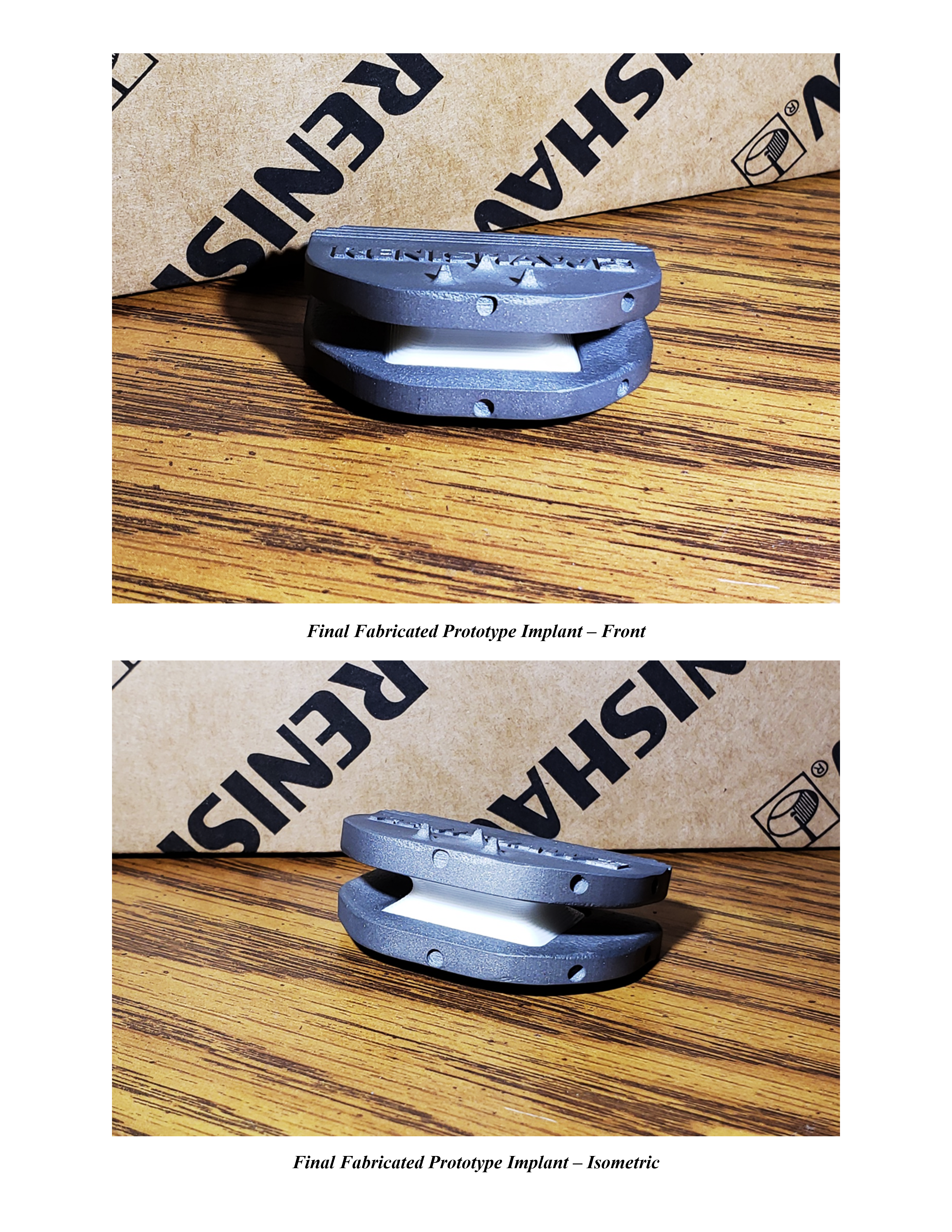
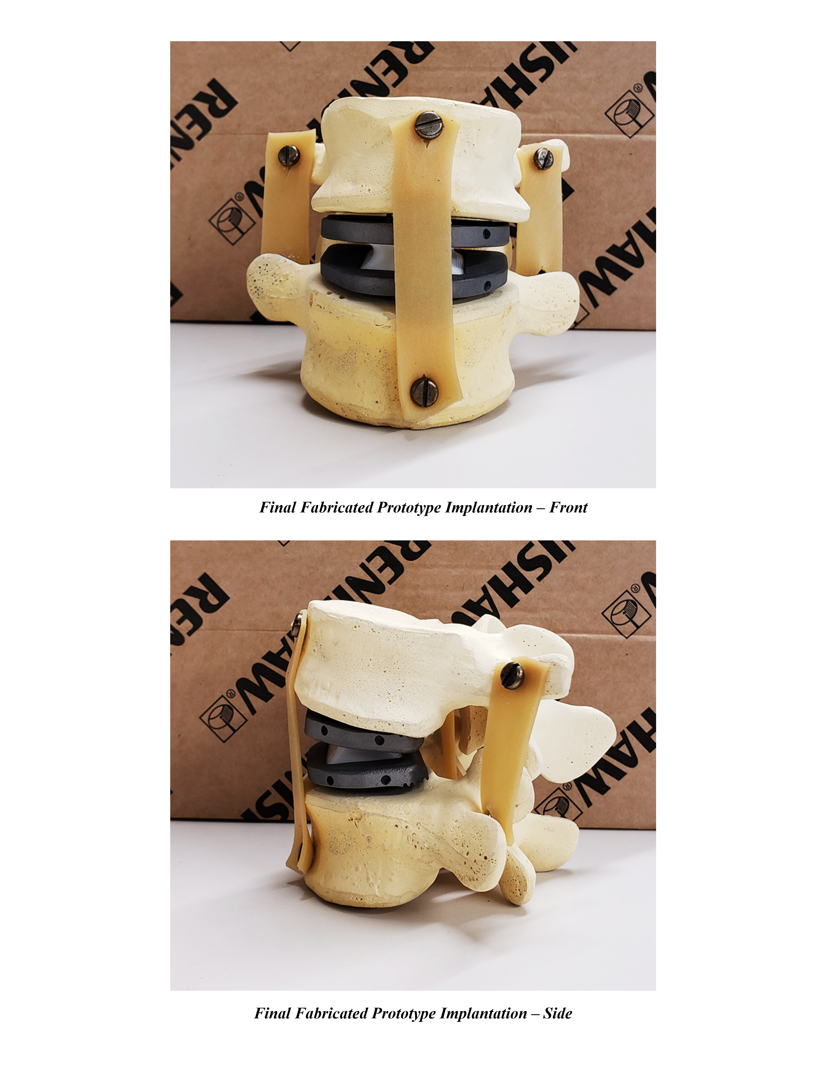
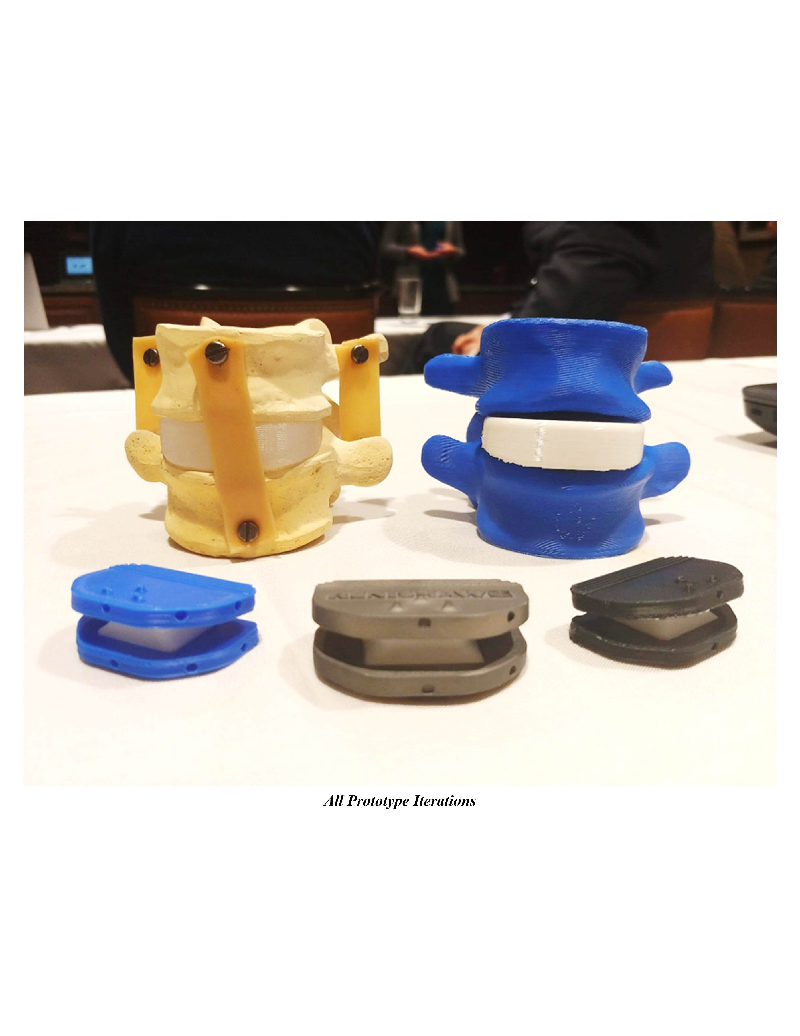
Ideation for this project began with identifying a joint in the body and an associating disease that required an implant solution. We chose to conduct extensive research through literature reviews on osteoarthritis in the spine and redesign a total disc replacement (TDR) implant. We were interested in creating an implant for the sufferers of Degenerative Disc Disease in order to grant them a better quality of life and allow them to perform normal daily activities that were taken away from them. Once the implant and joint were chosen, we were then tasked with identifying the limitations of the TDR implants currently on the market. During this process, we conducted further literature reviews and watched TDR surgeries to identify any limitations of the implant during the surgical procedure. As part of the preclinical testing stage, we created a CAD prototype of our redesigned implant and virtually implanted it into CT scanned bone for the L4/L5 vertebrae. FEA of the implant in the bone was completed in order to analyze the resulting force and stress distributions. The FEA results were then compared to data collected during motion capture for current implants and normal activities such as climbing stairs. Our final implant design was then 3D printed in Titanium by Renishaw plc. The artificial disc was 3D printed in Ninjaflex to demonstrate the motion of the implant.
Design Description:
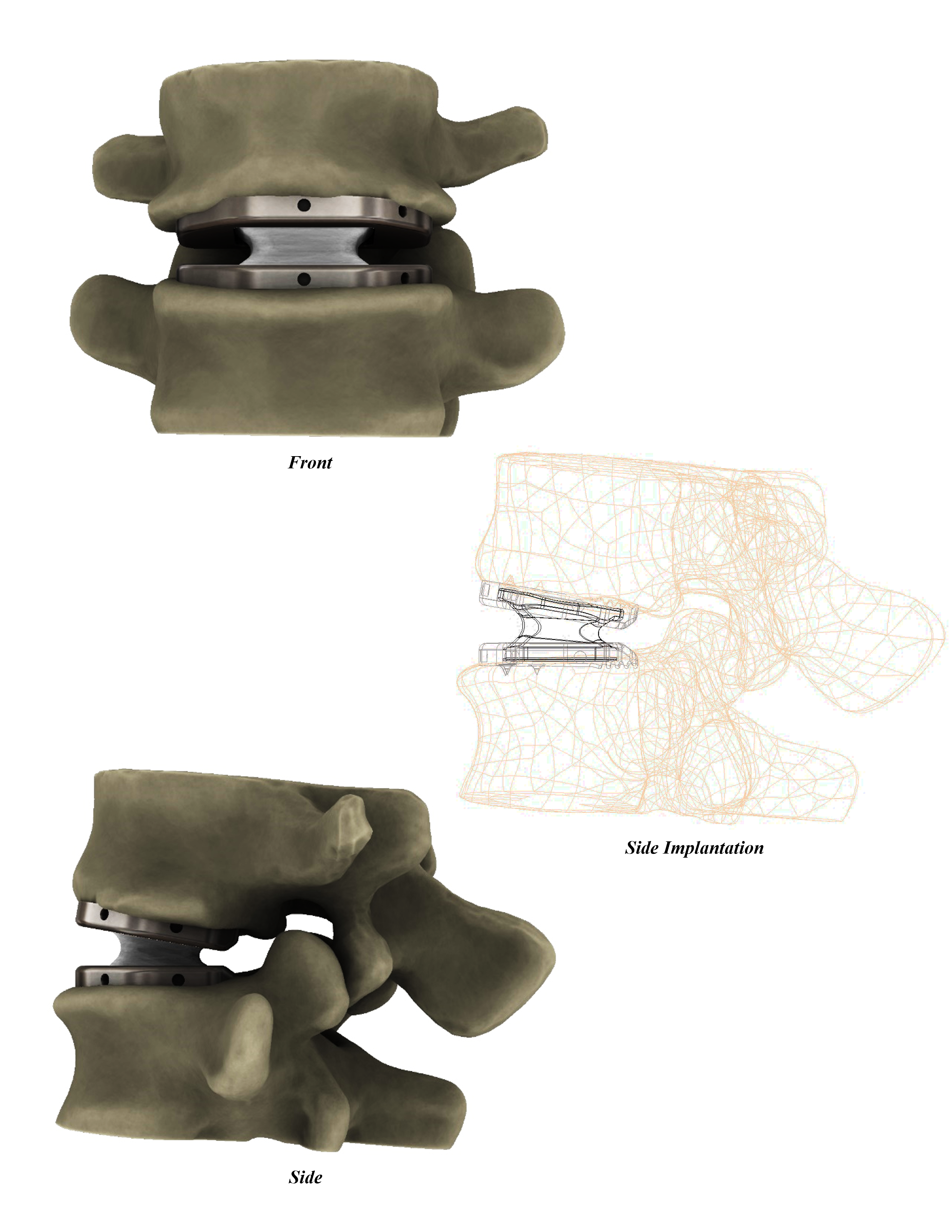
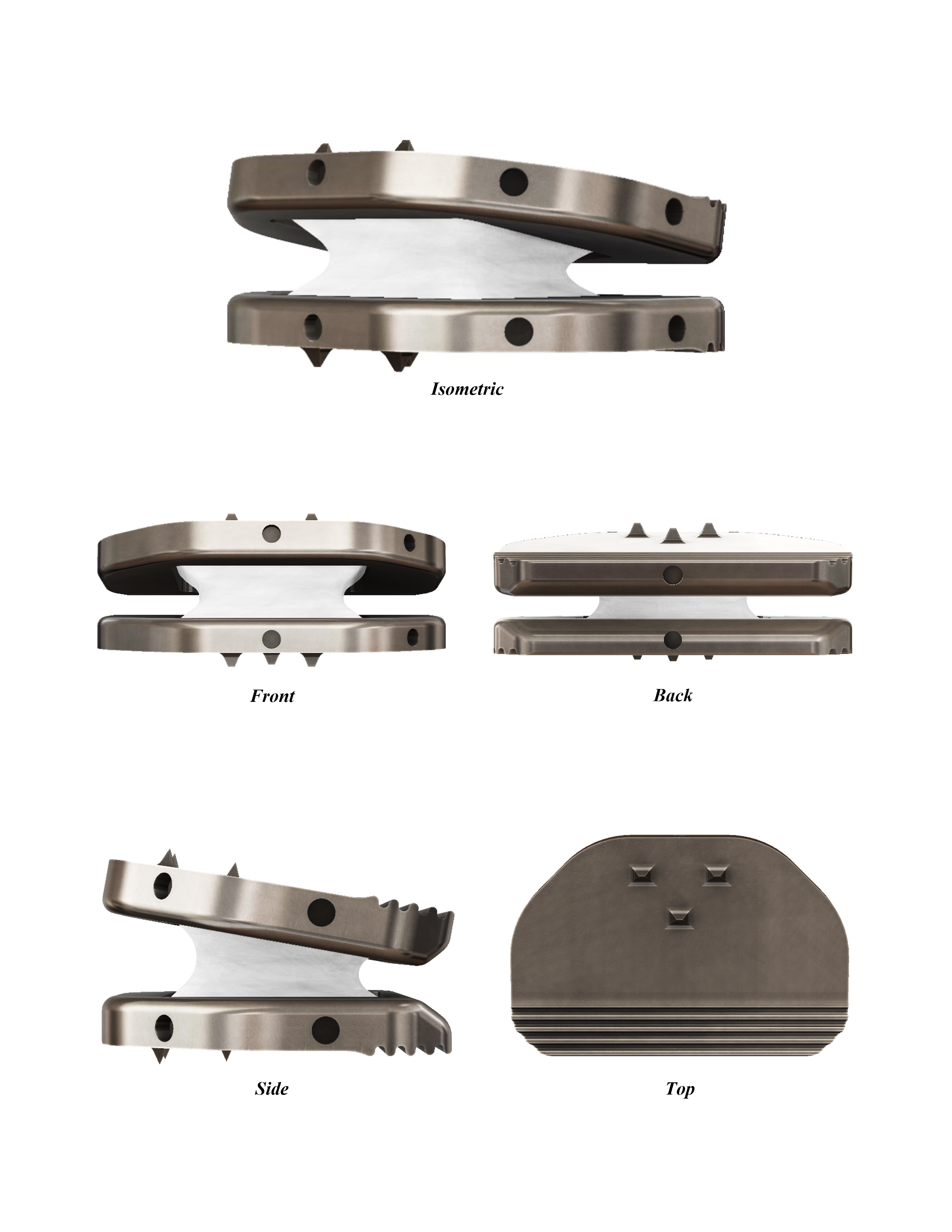
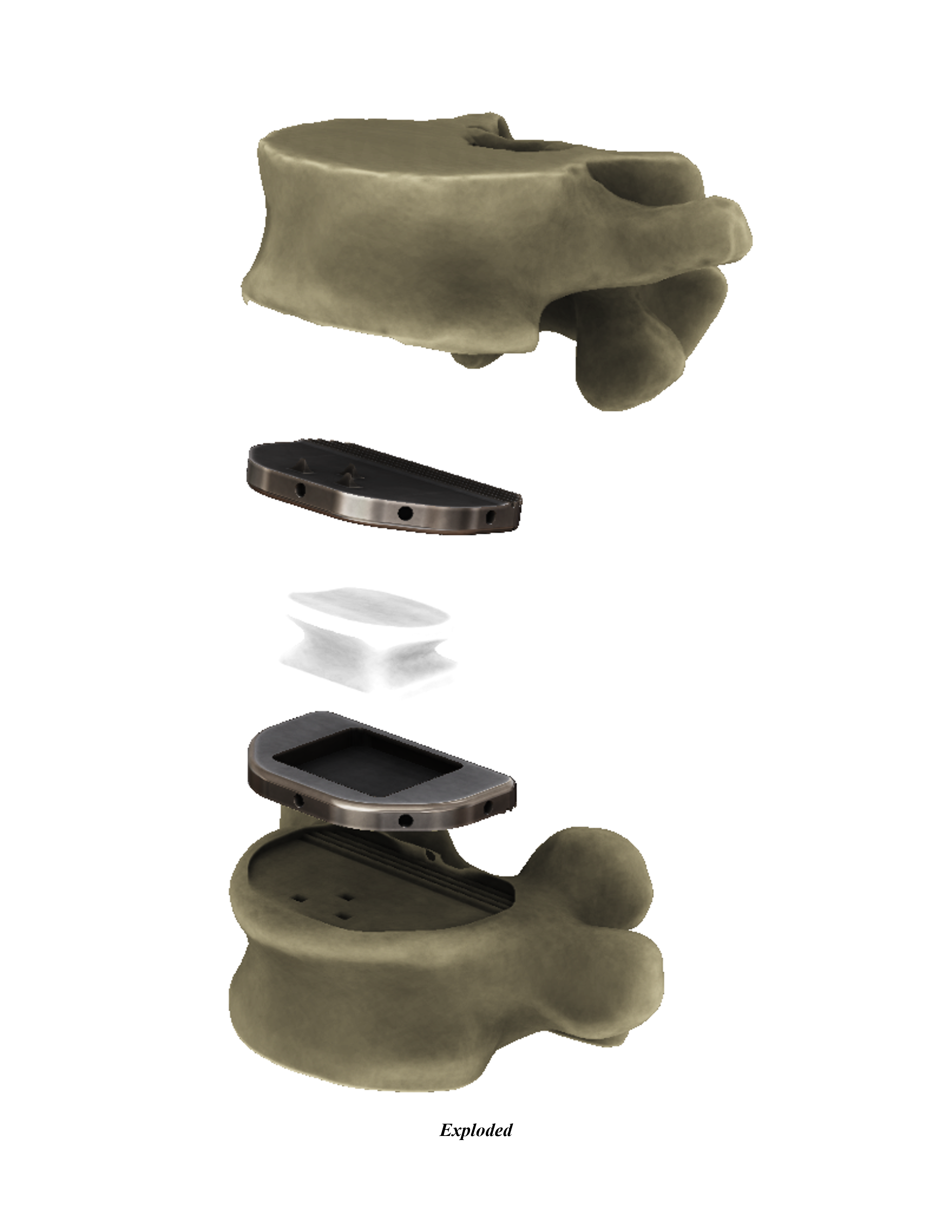
We developed a hybrid constrained lumbar L4/L5 TDR implant for lateral surgical implantation and with a ‘flexible fusion’ design. The ‘flexible fusion’ design was inspired during our interview process with various orthopedic surgeons. Upon asking their opinions of TDR implants, a common theme arose that orthopedic surgeons prefer fusion surgeries. They shared that fusion surgeries are more reliable even though they limit the patient’s overall range of motion. We then decided that we wanted to develop a TDR implant that would mimic the fixation of the fusion surgery while increasing the patient’s range of motion. This is where the ‘flexible fusion’ design was born. We decided to follow the success of previous implants in using a semi-constrained, metal-on-polymer design for our TDR implant. Inspiration was drawn from the general shape of existing implants (ActivL and ProDisc-L) as manufacturing techniques are likely standardized. The novelty of our design came from the surgical technique and shape of our artificial disc. Our TDR implant was comprised of 3 components: the superior endplate, inferior endplate, and artificial disc. Our endplates were designed to be manufactured from cobalt chromium and the artificial disc from ultra-high molecular weight polyethylene. We designed our implant for a lateral surgical approach which was based on existing techniques for spinal fusion procedures. Our implant was to be inserted by press-fitting the spikes located on each of the endplates into their respective vertebrae: the superior end of the superior endplate into the L4 vertebrae and the inferior end of the inferior endplate into the L5 vertebrae. These spikes would increase bone fixation while the sinusoidal pattern at the backend of the endplates would promote bone growth on the endplate surface. Our implant design was a hybrid between partially and fully constrained implants since both ends of the artificial disc were fixed into the endplates, however, the superior connection was free to rotate. This hybrid design was accomplished through the addition of endplate trenches. The endplate trenches are the location where the disc contacts the endplate. The inferior endplate had a rectangular trench for a snap-fit integration with the artificial disc while the superior endplate had a semicircular trench to allow disc rotation. This trench design in conjunction with our novel artificial disc geometry allowed our implant to achieve the ‘flexible fusion’ functionality we set out to achieve. Through our artificial disc’s unique geometry, it was able to restore the joint kinematics while limiting frictional wear during rotation. Instead of excessive wear occurring at the surface of the disc as in traditional means, our disc dissipated all forces through its core. Through the disc’s ‘X’ shape geometry, it was able to function as a shock absorber while allowing responsive behavior to the twisting and bending of the spine. The shape of each end of the disc (superior and inferior) corresponded to the geometry of the endplate trench that it articulated upon. Additionally, our disc incorporated the 10 degrees of the lordotic curve in the spine to further improve the patient’s range of motion.

University:
University of Illinois at Urbana-Champaign
Program:
B.S. in Mechanical Science and Engineering
Course:
ME 598: Orthopedic Biomechanics
Experience Level:
Senior Year
Project Duration:
4 Months
Valentino Ivan Wilson
Chicago, IL
