Valentino Ivan Wilson
Total Knee Arthroplasty Implant
Project Description:
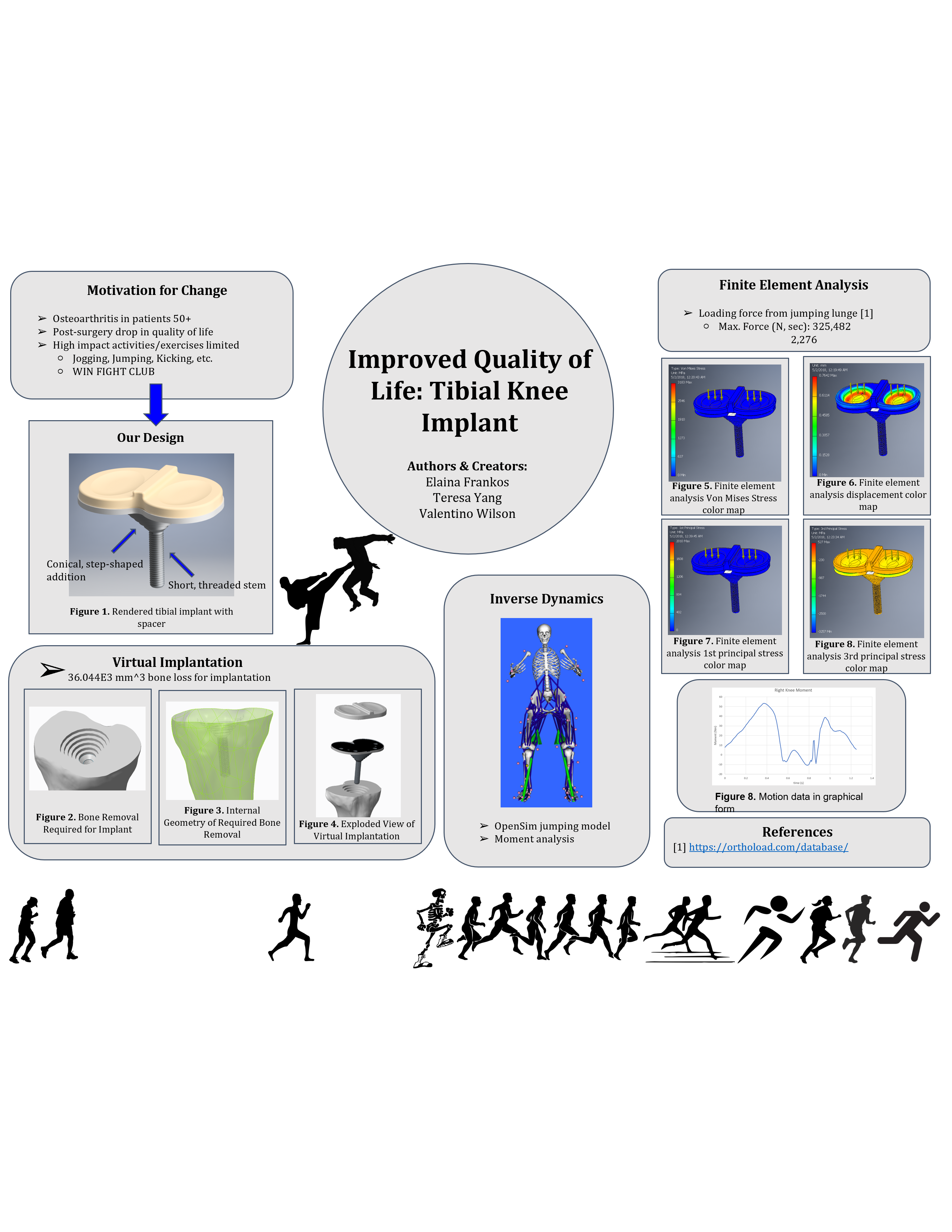
This group project tasked us with researching a clinical problem that required an implant solution at a specific joint in the body. We were then asked to propose a redesigned implant of our choosing that addressed underlying issues in the implant’s current design. The project was divided into three parts: implant proposal, design proposal, and a poster session. My group consisted of three students and my role in the project, aside from normal group member tasks, was creating all CAD models, final assembly, and corresponding GD&T engineering drawings. I was also responsible for conducting the virtual implantation and inverse dynamic calculations for our implant. All CAD was conducted in SolidWorks.
Design Procedure:
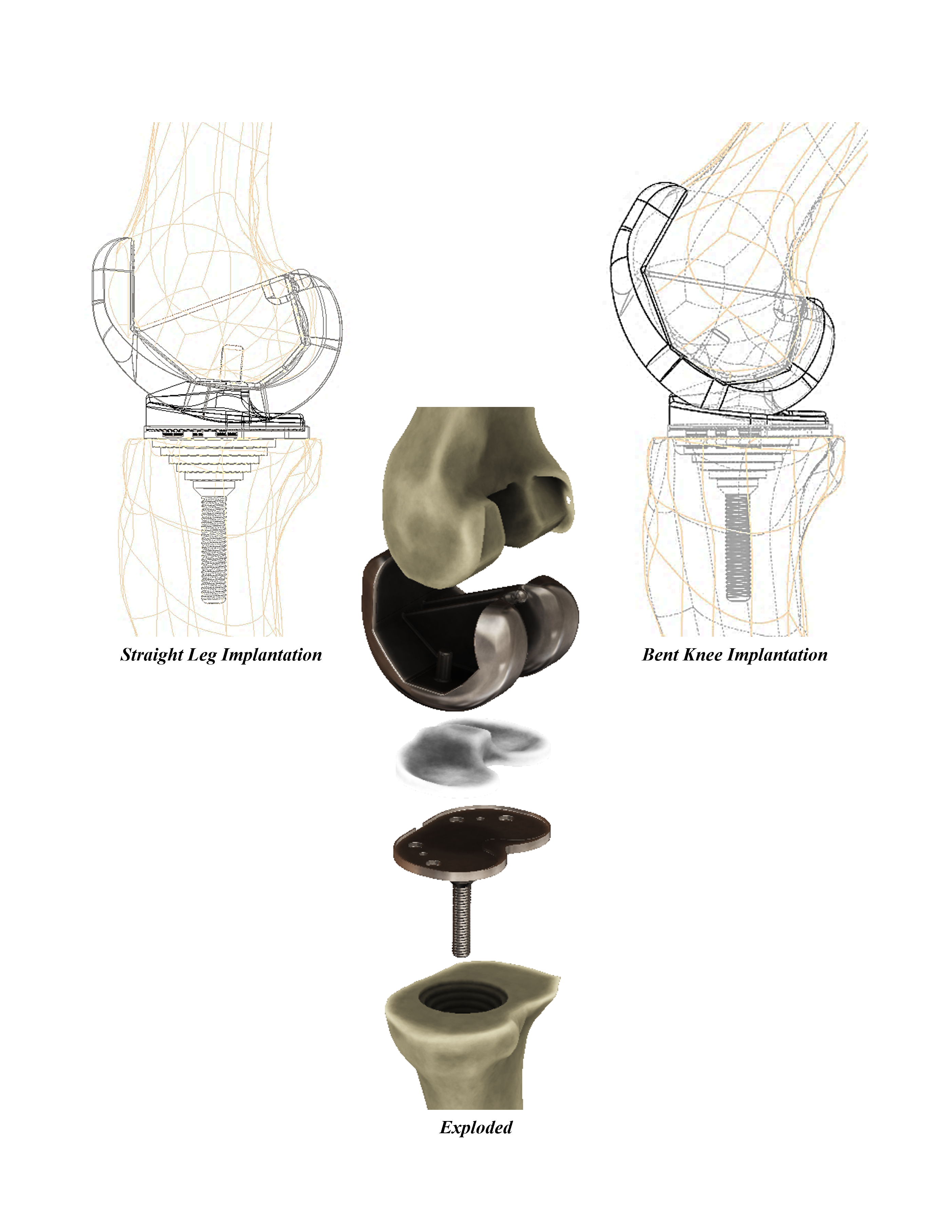
Ideation for this project began with determining a disease and joint of the body. We chose to conduct extensive research through literature reviews into knee osteoarthritis and redesign a total knee arthroplasty implant. We then determined our target user group and the criteria for which we wanted to redesign the implant. We decided to target the younger end of the spectrum (under 50 years of age) with the goal of reducing common restrictions on high-stress activities such as jogging, contact sports, or other sports that require twisting and pivoting. Our design goals for developing an implant that would allow for high-stress activities began with first determining a way to spread the forces from the implant to a wider surface area in the bone. We then wanted to use modern biocompatible surfaces instead of bone cement to affix the implant into the bone. Lastly, we wanted to replace the standard polyethylene spacer with a higher endurance material and increase its dynamic capability. A prototype assembly of our redesigned implant was created in CAD, and 3D printed in PLA plastic using Fused Deposition Modeling. Our final CAD implant was then virtually implanted into CT scanned bone for the femur and tibia. After virtual implantation occurred, FEA of the implant in the bone was completed in order to analyze the resulting force and stress distributions. The FEA results under normal loading conditions were then compared to data collected during motion capture when kicking a dummy (representative of martial arts or self-defense activities). Lastly, inverse dynamics of our chosen joint were completed, and GD&T engineering drawings of our final implant were created.
Design Description:
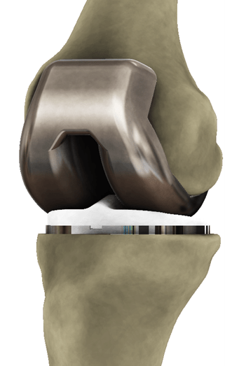
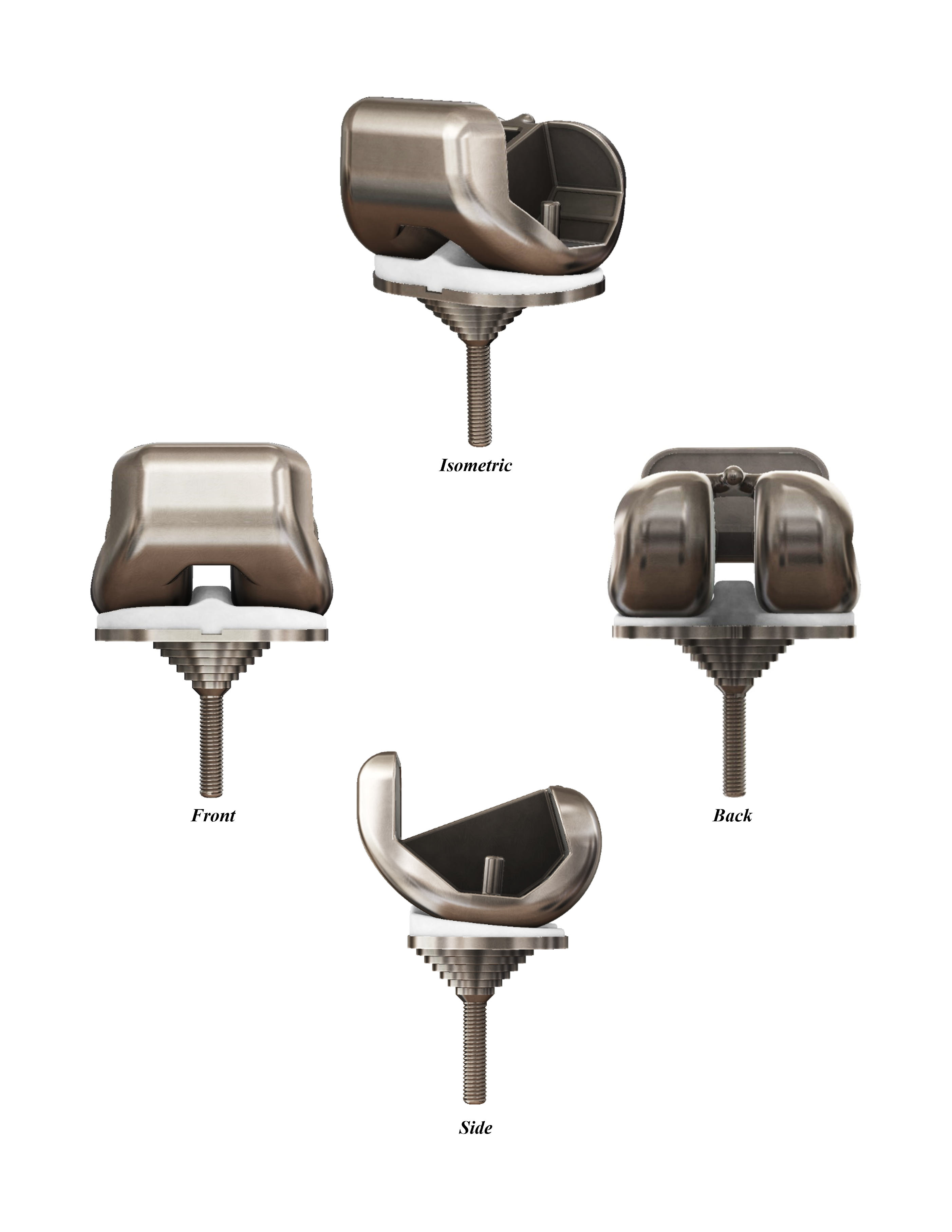
Our posterior stabilized knee implant consisted of two parts, the tibial and femoral components. The femoral component contained minimal design changes, while the tibial component contained all new modifications. The tibial component was comprised of three parts: the stem, tibial tray, and spacer. The stem is the part of the implant drilled into the bone and is responsible for proper fixation. The tibial tray is located proximally from the stem and houses the spacer. The spacer is a common plastic piece that cushions the joint and allows for the femoral cap to glide. The stem for our implant was designed to be shorter in length and completely threaded. The threads on the stem reduced the risk of unwanted displacement or rotation of the implant when subjected to strong impacts such as pivoting. Its combination of threaded and press-fit insertion into the bone also increased its likelihood for successful bone integration without the use of toxic bone cement which runs the risk of harming surrounding osseous tissue. A shorter stem length was chosen to combat possible stress shielding within the bone since we found that the degree of stress shielding is proportional to the length of the stem. To further reduce the stress concentration of the stem, our stem design has a rounded tip as well as a conical, step-shaped addition beneath the tibial tray. This step-shaped design adds a larger surface area for the compressive stress to be spread. We decided that our implant would be manufactured from a biomaterial made of tantalum called Trabecular Metal. This material has a low modulus of elasticity (similar to bone) resulting in a reduction in stress shielding and a more “natural” load response. It also has a high porosity of up to 80%, which allows for excellent bone ingrowth. The spacer for our implant featured a cam and post pairing with the femoral component that prevented the femoral cap from translating completely out of the knee joint and causing joint dislocation. Additionally, the cam was designed with a triangular cross-section in order to ensure that the femoral cap glided across the spacer in a fashion that more closely replicated the natural motion of the knee. The implant thickness was also increased to provide deeper grooves for femoral cap gliding. We chose to manufacture our implant spacer from Teflon Polytetrafluoroethylene (PTFE) as it is a biocompatible material that is chemically inert, non-toxic, has good tensile strength, features zero moisture absorption, has a low coefficient of friction, and is also resistant to high heat.
University:
University of Illinois at Urbana-Champaign
Program:
B.S. in Mechanical Science and Engineering
Course:
ME 481: Whole Body Musculoskeletal Biomechanics
Experience Level:
Junior Year
Project Duration:
3 Months

Valentino Ivan Wilson
Chicago, IL
